Fluorescence microscopy is a powerful imaging technique based on the ability of certain materials to emit visible light after being exposed to a specific wavelength of light. This emission occurs when a substance absorbs energy and then re-emits it as light. In fluorescence microscopy, the sample being observed can either be naturally fluorescent or treated with special fluorescent substances that allow certain components of the sample to glow under specific lighting conditions.
In traditional microscopy, the light you see through the eyepiece is simply the light that has passed through or been reflected off the sample. However, fluorescence microscopy works quite differently. In this technique, the light seen in the eyepiece is not the original light emitted by the microscope’s light source. Instead, it is light emitted by the specimen itself after being excited by a specific wavelength of light. To achieve this, a high-intensity light source is required.
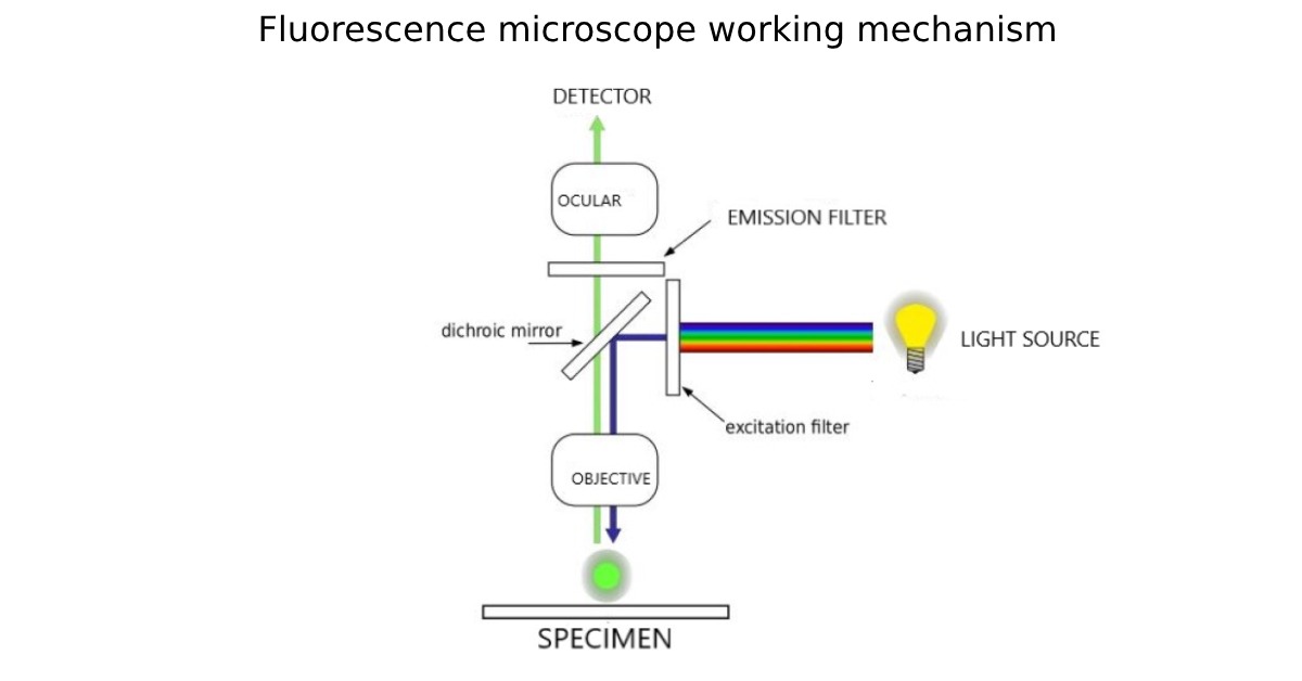
This light is first passed through a dichroic filter cube, which contains a fluorescence bandpass excitation filter. This filter allows only light of specific wavelengths to pass through, directing it to the sample. When the sample absorbs this energy, it undergoes a transformation and re-emits light of a longer wavelength, which is then collected by the microscope’s objective lens. Any light that reflects back into the microscope’s objective is filtered out by an emission filter before the fluorescence light is allowed to pass through. This emitted fluorescence is what we observe in the microscope’s eyepieces, producing a bright, colorful image of the sample.
There are two primary methods for illumination in fluorescence microscopy: dark-ground illumination and epifluorescence illumination. Dark-ground illumination separates the excitation light and the emitted fluorescence light, but epifluorescence is more advanced. In epifluorescence microscopy, the excitation light is directed onto the specimen from above using the same objective lens that also collects the emitted light. This approach allows the excitation light to travel directly through the sample, which results in a better signal-to-noise ratio, as most of the excitation light simply passes through the specimen. Only the emitted fluorescence, along with any reflected excitation light, is collected by the objective lens and sent to the detector, making the image clearer and brighter.
To further enhance the quality of the fluorescence image, an additional filter is often placed between the objective lens and the detector. This filter ensures that only the emitted fluorescence light is passed through, blocking out any remaining excitation light. This combination of techniques leads to better image quality and more precise observations of fluorescent materials within the specimen.
Epifluorescence Microscopy in Life Sciences
Epifluorescence microscopy (EFM) is increasingly used in biological and medical research due to its ability to offer high specificity in imaging. This method has revolutionized the study of cellular structures, allowing scientists to identify and study specific components of cells with remarkable precision. For example, EFM is widely used to examine diseases, identify impurities, and study cellular mechanisms. Researchers can label specific proteins or organelles with fluorescent markers, allowing them to track these components in live cells or tissues.
One of the key advantages of EFM is its ability to capture depth-discriminated, or optical sectioned, images of samples. Modern fluorescence microscopes, such as those equipped with systems like the ApoTome, offer the capability to produce optical slices of fluorescent samples. These images are not only clearer but also offer increased contrast and improved optical resolution in the axial direction (the Z-axis). This feature helps researchers to obtain more detailed three-dimensional views of the specimen, making it easier to study structures that may not be apparent in a conventional two-dimensional view.
Conclusion
Epifluorescence microscopy is an indispensable tool in modern biological and medical research. It enables scientists to observe specific cellular components with exceptional precision and clarity. By using specific fluorescent markers and high-intensity light sources, researchers can study a wide range of biological phenomena, from protein interactions to disease progression. With technological advancements such as optical sectioning and improved filters, epifluorescence microscopy continues to enhance the quality and scope of scientific discovery. As the technique becomes more advanced, its applications are likely to expand even further, offering new insights into the microscopic world of living organisms.


