
Dermatology, the branch of medicine dealing with skin, hair, and nails, has significantly evolved with advancements in technology. Among the tools transforming this field, microscopes play a vital role in diagnosing conditions, conducting research, and advancing therapeutic methods. With their ability to magnify and analyze the intricate structures of skin and its components, microscopes are indispensable in dermatological practice and research.
Why Microscopes Are Crucial in Dermatology
The human skin, being the largest organ, is a complex structure with multiple layers, each serving unique functions. Analyzing these layers with precision is essential to understanding skin conditions. Microscopes provide dermatologists with detailed insights into the cellular and molecular composition of the skin, helping in accurate diagnosis and effective treatment planning.
Types of Microscopes Used in Dermatology
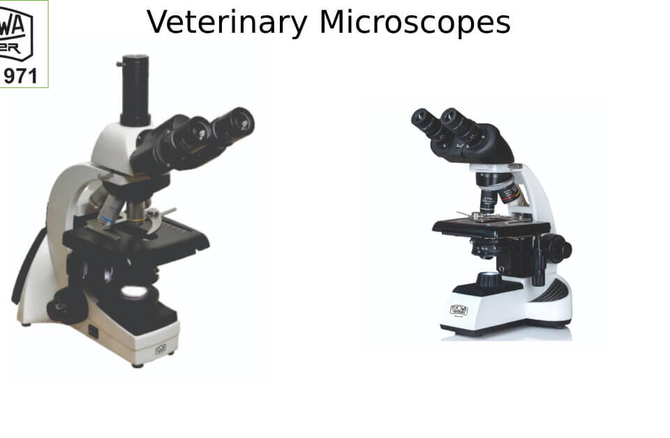
1. Light Microscopes
- Applications: Commonly used for routine histopathological analysis of skin biopsies.
- Utility: Helps in identifying skin diseases such as psoriasis, eczema, or melanoma by examining tissue samples.
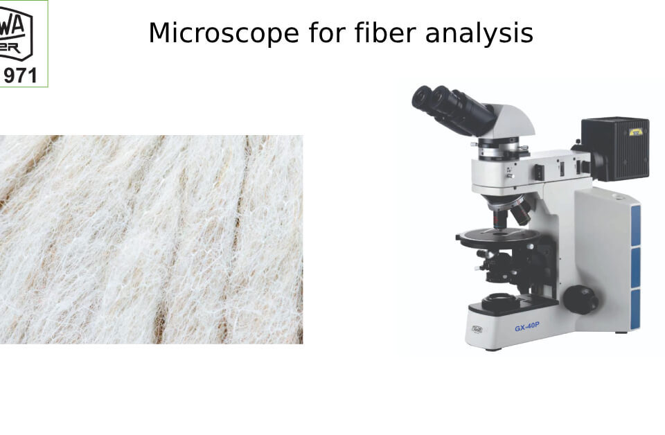
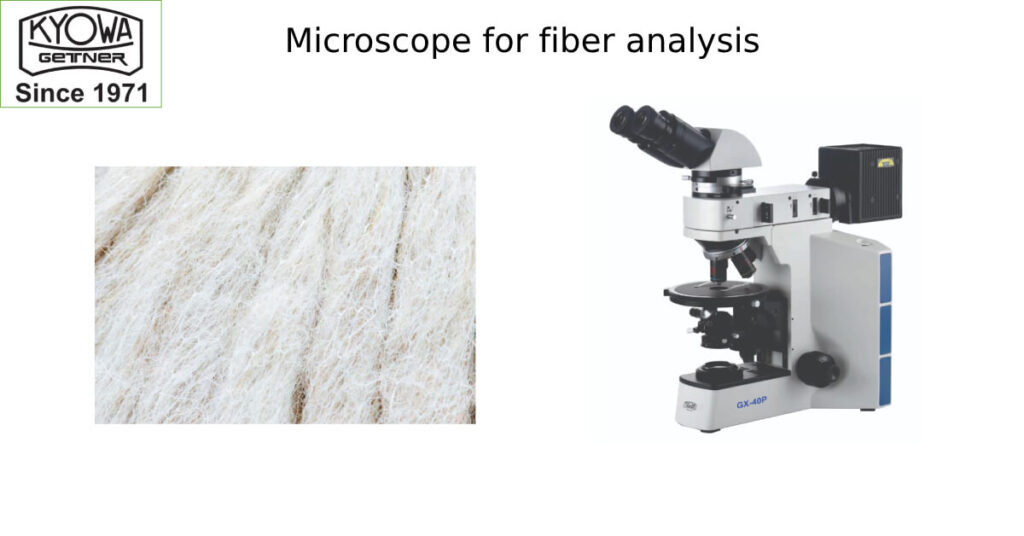
2. Polarizing Microscopes
- Applications: Used to study birefringent materials such as hair shafts and crystals in skin diseases like gout.
- Utility: Effective for detecting fungal infections and foreign bodies.
3. Confocal Microscopes
- Applications: Provides non-invasive imaging of the skin in vivo, enabling real-time analysis of skin layers.
- Utility: Popular for diagnosing skin cancers, such as melanoma, and for monitoring treatment efficacy.
4. Fluorescence Microscopes
- Applications: Used for immunofluorescence studies, where antibodies tagged with fluorescent dyes bind to specific skin proteins.
- Utility: Essential for diagnosing autoimmune blistering diseases like pemphigus vulgaris or lupus.
5. Electron Microscopes (SEM and TEM)
- Applications: Offers ultra-high magnification to study skin cells and structures at the nanoscale.
- Utility: Crucial for research into genetic skin disorders, viral infections, and complex dermatological conditions.
6. Digital Dermatoscopes
- Applications: Used for capturing high-resolution images of moles, lesions, or skin anomalies.
- Utility: Assists in early detection of melanoma and aids in teledermatology consultations.
Applications of Microscope for Dermatology
1. Skin Cancer Diagnosis
- Microscope for dermatology are essential in identifying cancerous cells in skin biopsies.
- Confocal microscopy enables real-time, non-invasive imaging for early detection of melanoma.
2. Autoimmune and Inflammatory Diseases
- Direct immunofluorescence (DIF) microscopy helps in diagnosing autoimmune diseases such as lupus, pemphigus, and vasculitis.
- Polarizing microscopes aid in identifying crystals in conditions like dermatomyositis.
3. Fungal and Bacterial Infections
- Microscopic examination of skin scrapings and biopsies allows dermatologists to identify fungi (e.g., ringworm, candida) and bacteria (e.g., leprosy).
4. Hair and Scalp Analysis
- Trichology, the study of hair and scalp disorders, heavily relies on microscopes to identify conditions like alopecia, fungal infections, and hair shaft abnormalities.
5. Research and Development
- Microscopes are integral in dermatological research, such as studying the effects of skincare products, anti-aging formulations, and wound healing treatments.
- Electron microscopy allows researchers to analyze cellular structures and molecular interactions.
6. Aesthetic Dermatology
- Microscopes are used to study skin rejuvenation procedures, laser treatments, and cosmetic enhancements, ensuring safety and efficacy.
Advancements in Dermatological Microscopy
1. Multiphoton Microscopy
A cutting-edge technique that uses near-infrared light to image deeper layers of the skin. It’s especially useful for studying skin aging and collagen remodeling.
2. Artificial Intelligence (AI) Integration
AI-powered microscopes can analyze skin samples more efficiently, assisting in faster diagnosis and predictive analytics for treatment outcomes.
3. Portable Microscopes for Teledermatology
Compact and digital microscopes allow dermatologists to diagnose conditions remotely, bridging the gap in dermatological care in rural areas.
The Future of Microscopy in Dermatology
The integration of advanced microscopy techniques, such as super-resolution microscopy and AI-driven imaging systems, will revolutionize the field of dermatology. Non-invasive diagnostic methods and enhanced imaging capabilities will make skin analysis faster, safer, and more precise.
Conclusion
Microscopes are the backbone of dermatological diagnosis and research. From identifying cancerous lesions to studying autoimmune diseases, these tools have transformed the way skin health is understood and treated. As technology continues to evolve, the applications of microscopes in dermatology will only expand, paving the way for improved patient care and groundbreaking discoveries.
For dermatologists and researchers seeking reliable and advanced microscopy solutions, “KYOWA-GETNER“, offers cutting-edge tools tailored to dermatology’s unique needs. Unlock the potential of microscope for dermatology and redefine your dermatological practice with precision and innovation.

