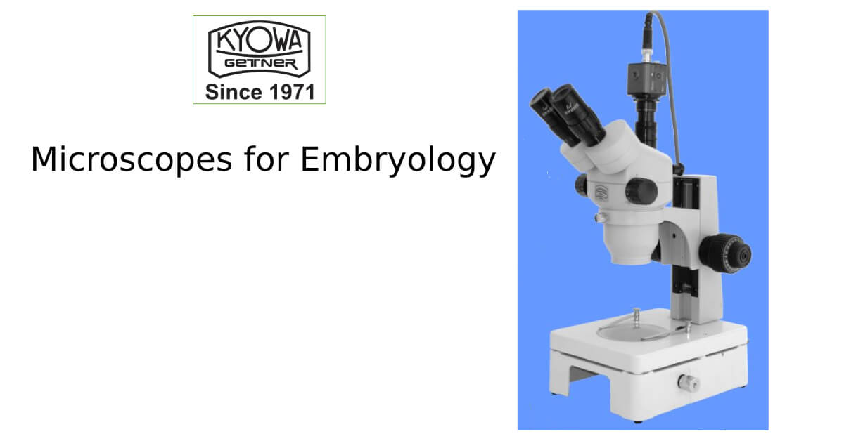
Embryology, the study of the development of embryos, is a fascinating and critical field of biology that has provided profound insights into the mechanisms of life. From fertilization to the formation of organs and tissues, embryological development is a complex and highly regulated process. To explore and understand these processes, scientists rely on a variety of sophisticated tools, and perhaps none more important than the microscope.
In this blog post, we will explore the types of microscopes used in embryology, their applications, and why they are indispensable in the study of early development.
The Role of Microscopy in Embryology
Embryology is inherently microscopic. Whether you’re studying the development of a single-celled zygote or the intricate differentiation of tissues in a multi-cellular embryo, understanding these processes requires a high degree of magnification and precision. Microscopes allow embryologists to observe cellular structures, developmental stages, and molecular processes that would otherwise be invisible to the naked eye. These observations are crucial for understanding how cells divide, migrate, and differentiate to form tissues and organs.
Additionally, advanced microscopes help researchers observe the effects of genetic mutations, environmental factors, and other variables on embryonic development, providing invaluable insights into everything from fertility and developmental disorders to evolutionary biology.
Types of Microscopes Used in Embryology
Light Microscopes
The light microscope, or compound microscope, is the most commonly used tool in embryology. It works by passing visible light through a specimen, which is then magnified and projected through a series of lenses. Light microscopes can be equipped with different types of objectives and condensers, allowing for a range of magnifications from 40x to 1000x.
Applications in Embryology:
- Cell Division: Light microscopes allow embryologists to observe mitosis and meiosis in developing embryos. For example, watching the zygote undergo cleavage (early cell divisions) is a fundamental process in embryology.
- Organogenesis: As organs begin to form and differentiate, light microscopes help visualize these processes at a cellular level.
- Live Imaging: Some advanced light microscopes allow the observation of live embryos in real-time, enabling researchers to track developmental processes such as cell migration, differentiation, and morphogenesis.
Phase Contrast Microscopes
Phase contrast microscopy is an enhancement of the basic light microscope. It uses differences in the refractive index of the different parts of the specimen to produce higher contrast images without staining. This is especially useful for observing live, unstained embryos and cells.
Applications in Embryology:
- Live Embryo Observation: Phase contrast microscopes are widely used in embryology to observe live embryos without damaging them through staining. Researchers can track the development of embryos in real-time, capturing dynamic processes like gastrulation, organ formation, and neuronal development.
- Cytoskeletal Studies: The phase contrast technique allows embryologists to observe structures such as the mitotic spindle and other cytoskeletal components as they form during cell division.
Fluorescence Microscopes
Fluorescence microscopy takes advantage of fluorescence to create highly detailed images of specimens. This technique involves labeling specific molecules within the embryo with fluorescent dyes or proteins, which then emit light when excited by a particular wavelength. This provides both high contrast and resolution, revealing structures at the molecular and cellular levels.
Applications in Embryology:
- Gene Expression: Fluorescence microscopy is ideal for studying gene expression patterns in embryos. By tagging specific genes with fluorescent markers, embryologists can observe where and when particular genes are activated during development.
- Protein Localization: This technique also allows for the localization of specific proteins within cells. For example, observing the role of transcription factors or signaling molecules during early developmental stages.
- Tracking Cell Lineage: By using fluorescence markers, scientists can trace the lineage of specific cells within an embryo and follow their developmental fate.
Confocal Microscopes
Confocal microscopy is an advanced form of fluorescence microscopy that uses lasers to focus on specific layers of a sample. It captures images at various depths of the embryo, creating highly detailed three-dimensional reconstructions. This is particularly useful for studying thick or multi-layered specimens.
Applications in Embryology:
- 3D Imaging of Development: Confocal microscopes can create detailed 3D images of developing embryos, allowing scientists to visualize complex structures such as the neural tube, heart, or vasculature as they form.
- Cellular Dynamics: Confocal microscopy can track the movement of individual cells during development, such as cell migration during gastrulation or epithelial-to-mesenchymal transition (EMT).
- Cell Interactions: It is particularly useful for studying interactions between different cell types within the developing embryo, such as those involved in tissue patterning.
Why Microscopes Are Indispensable in Embryology
Microscopes allow embryologists to probe the microscopic world of developing embryos in ways that are crucial for advancing our understanding of biology. From studying the molecular basis of developmental processes to investigating the effects of genetic mutations or environmental factors, microscopy provides the resolution, contrast, and depth of insight needed to explore the earliest stages of life.
In clinical embryology, microscopes are essential for techniques such as in vitro fertilization (IVF), where embryologists monitor the development of embryos outside the body. Similarly, the study of developmental disorders, such as congenital birth defects or stem cell therapy, relies heavily on advanced microscopy to understand cellular and molecular abnormalities.
Moreover, as microscopy technologies continue to evolve, researchers can now explore more detailed and dynamic processes, opening up new frontiers in understanding human development, regenerative medicine, and evolutionary biology.
Conclusion
Microscopes are the unsung heroes of embryology, providing a window into the minute details of life’s earliest stages. Whether using light microscopy for basic observation, fluorescence microscopy to track gene expression, or electron microscopy to explore cellular structures, these powerful tools enable scientists to uncover the mysteries of development with incredible precision. As the field of embryology continues to advance, the role of microscopes will remain central to unraveling the complexities of life’s origins and the processes that guide the development of organisms.


