
Histology, the study of microscopic tissue structures, plays a crucial role in medical diagnostics, research, and education. At the heart of histological studies are microscopes, which enable scientists and medical professionals to observe intricate tissue details at the cellular level. Choosing the right microscope is essential for accurate histological examination.
Types of Microscopes Used in Histology
1. Light Microscopes
Light microscopes are the most commonly used instruments in histology labs. They work by transmitting visible light through thinly sectioned tissue samples. The most common types include:
- Brightfield Microscopes: These are widely used for routine histology. They provide clear images of stained tissue sections, making them ideal for general pathology and anatomy studies.
- Phase Contrast Microscopes: Used for examining unstained or living tissue samples, phase contrast microscopes enhance contrast by exploiting differences in refractive index.
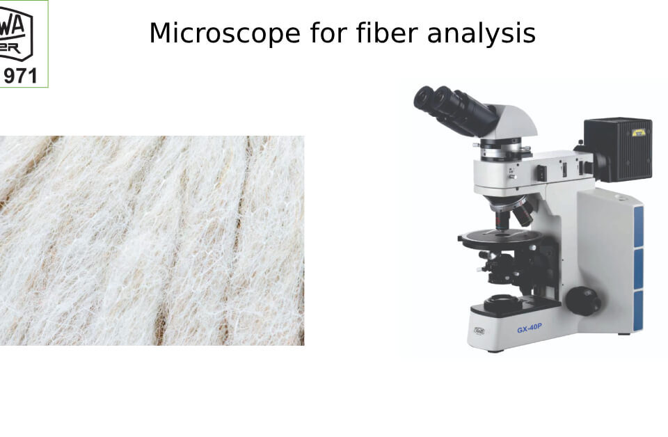
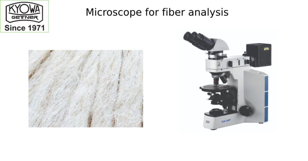
- Polarized Light Microscopes: These are useful in detecting birefringent structures, such as collagen fibers or crystalline deposits in tissues.
2. Fluorescence Microscopes
Fluorescence microscopy is a powerful tool in histology, especially for immunohistochemistry and molecular diagnostics. By tagging tissue components with fluorescent dyes, researchers can detect specific proteins, DNA sequences, and cellular markers.
3. Confocal Microscopes
Confocal microscopy provides high-resolution, 3D imaging of tissues by using laser scanning technology. It is widely used in histopathology, neuroscience, and cancer research to study cell structures with precision.
4. Electron Microscopes (EM)
For ultra-detailed tissue analysis, electron microscopes offer much higher magnification and resolution than light microscopes. There are two main types:
- Transmission Electron Microscopes (TEM): Provide high-resolution images of thin tissue sections, revealing organelles and intracellular structures.
- Scanning Electron Microscopes (SEM): Generate 3D images of tissue surfaces, ideal for studying tissue morphology.
Key Features to Consider in a Histology Microscope
When selecting a microscope for histological studies, consider the following features:
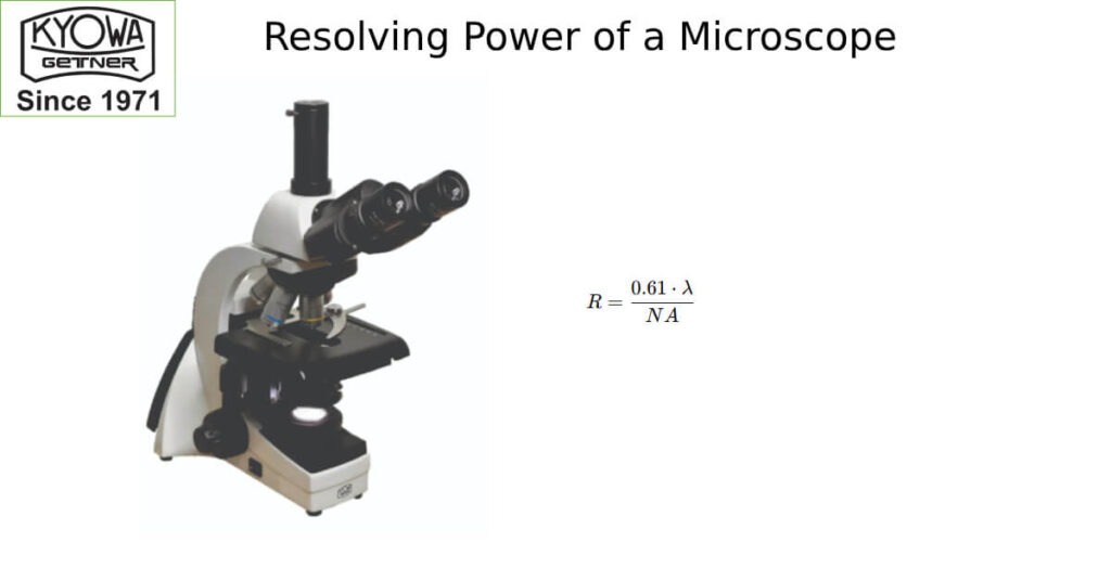
- Magnification & Resolution: Most histology applications require objectives ranging from 4x to 100x with oil immersion for high-resolution imaging.
- Optical Quality: High-quality achromatic, plan-achromatic, or apochromatic lenses ensure sharp, distortion-free images.
- Stage & Slide Holders: A mechanical stage with smooth movement is crucial for precise slide examination.
- Illumination System: LED or halogen illumination with adjustable brightness enhances visibility and contrast.
- Camera Integration: Digital microscopes with built-in cameras facilitate documentation, analysis, and remote collaboration.
Applications of Microscopes in Histology
Microscopes are indispensable in various histological applications, including:
- Medical Diagnostics: Pathologists use microscopes to detect abnormalities in tissue samples, aiding in disease diagnosis.
- Cancer Research: Identifying malignant cells and studying tumor microenvironments.
- Neuroscience: Investigating brain and nervous system tissues.
- Pharmaceutical Research: Evaluating tissue response to new drugs.
- Education & Training: Teaching students about tissue structures and disease pathology.
Conclusion
Microscopes are the foundation of histological studies, enabling researchers and medical professionals to explore the intricate details of tissues. Whether for routine laboratory work or advanced research, selecting the right microscope ensures accuracy, efficiency, and better diagnostic outcomes. With advancements in imaging technology, histology microscopes continue to evolve, providing even greater insights into cellular and molecular structures.
If you are looking for high-quality microscopes for histology, explore KYOWA-GETNER range of advanced biological and digital microscopes designed for precise tissue analysis.
