Phase contrast microscopy (PCM) is one of the most significant advancements in optical microscopy that allows researchers to observe transparent and living cells without the need for dyes or stains. Developed by Dutch scientist Frits Zernike in 1932, phase contrast microscopy has revolutionized the way we study biological specimens, providing high-contrast images of cellular structures and activities in their natural state. This blog will explore the working principle, advantages, and applications of phase contrast microscopy, demonstrating why it remains an essential tool in biological research and medical diagnostics.
What is Phase Contrast Microscopy?
Phase contrast microscopes work by converting phase differences in light passing through transparent specimens into visible light intensity differences. Unlike brightfield microscopy, which requires specimens to be stained to provide contrast, phase contrast microscopy enables the study of living cells without altering or killing them. This technique is particularly useful for examining living cells, microorganisms, and tissue cultures—all of which are often transparent and difficult to observe using traditional methods.
The main advantage of phase contrast microscopy is its ability to provide high-contrast images from colorless biological samples without the need for staining agents or other chemical preparations. This makes it especially valuable for long-term live-cell imaging, enabling scientists to track cellular processes like cell division, movement, and morphology.
How Does Phase Contrast Microscopy Work?
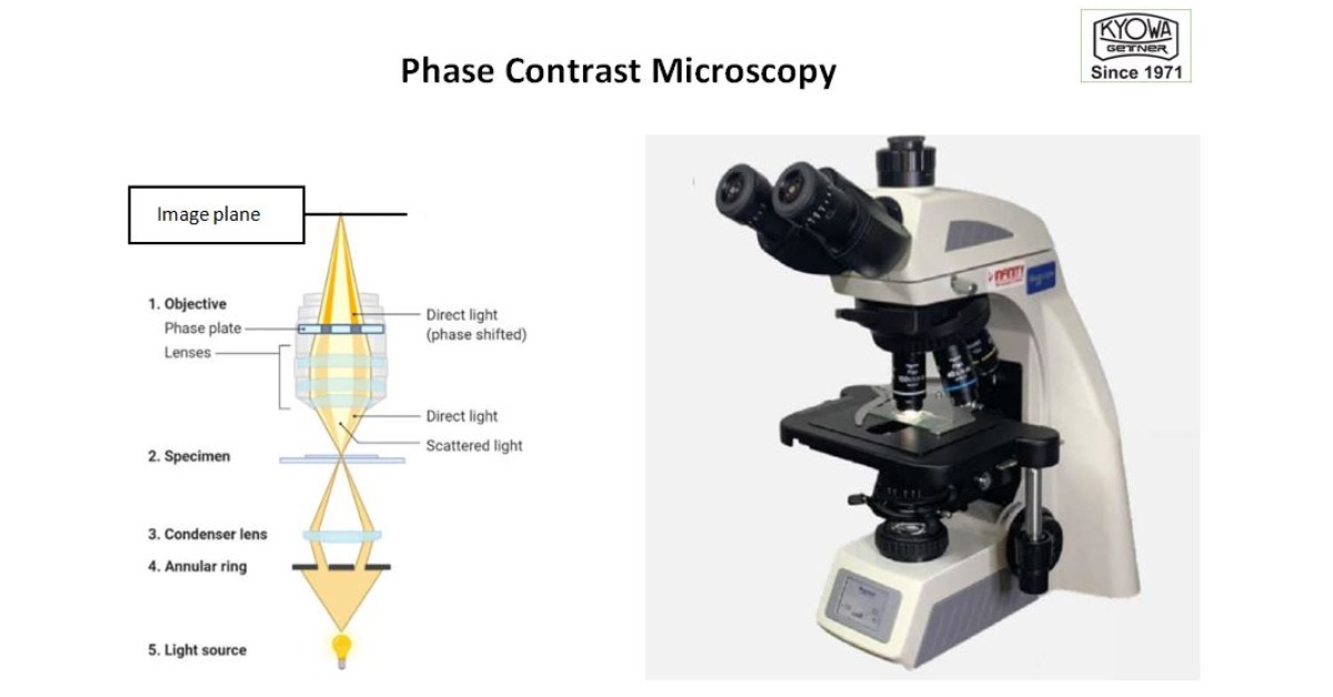
The key to phase contrast microscopy is the way it manipulates light waves passing through a sample. Here’s a step-by-step breakdown of the working principle of PCM:
- Illumination: Light from the microscope’s illuminator passes through a condenser, focusing the light onto the specimen.
- Phase Shifting: As the light travels through the sample, the different components of the sample—such as cell membranes, organelles, and cytoplasm—affect the light waves by altering their phase (the timing of the light waves). Areas with different refractive indices (e.g., denser structures like the nucleus) cause greater phase shifts.
- Interference: Phase contrast microscopes contain specialized optical components (such as a phase plate and phase ring) that convert phase differences into variations in light intensity. This interference between light waves results in enhanced contrast in the final image.
- Image Formation: The interference patterns create high-contrast images, allowing structures like cell nuclei, vacuoles, and other fine details to be visible, even if they are otherwise transparent.
Advantages of Phase Contrast Microscopy
Phase contrast microscopy has several key benefits that make it a powerful tool for studying biological samples:
- Live Cell Imaging: Phase contrast microscopy allows for the study of living cells in real time, without the need for chemical staining. This is ideal for observing cellular processes like cell division, migration, and intracellular movement in live cultures.
- No Staining Required: Unlike techniques like fluorescence microscopy or brightfield microscopy, phase contrast microscopy doesn’t require any staining or preparation, preserving the natural state of the sample. This is crucial when working with delicate, living samples like embryos, tissues, or microorganisms.
- High Contrast for Transparent Samples: Biological specimens, such as microorganisms, cellular components, and embryos, are often transparent and lack natural contrast. Phase contrast enhances the visibility of these structures by amplifying refractive index differences, allowing for detailed observation of subcellular features.
- Real-Time Monitoring: The ability to observe live, dynamic processes is one of the greatest advantages of phase contrast microscopy. Researchers can track changes in living cells over time, making it an essential tool for studying cellular behavior and disease processes.
- Ease of Use and Affordability: PCM can be performed using relatively affordable, standard optical microscopes, making it accessible for many laboratories. It requires only minimal setup adjustments, such as installing a phase contrast objective and condenser, making it a user-friendly technique.
Applications of Phase Contrast Microscopy
Phase contrast microscopy has a wide range of applications in various fields, from cell biology to microbiology. Below are some of the most prominent areas where this technique is used:
- Cell Biology: PCM is indispensable for studying the inner workings of cells. Researchers use it to observe cell division, morphological changes, and intracellular structures in living cells, making it an invaluable tool in cell culture and stem cell research.
- Microbiology: Phase contrast is widely used to study microorganisms such as bacteria, fungi, and protozoa. It allows microbiologists to observe the morphology and behavior of these organisms without the need for chemical staining, which can be harmful to delicate specimens.
- Developmental Biology: In developmental biology, PCM is crucial for tracking embryonic development. It allows scientists to study processes such as embryo growth, cell differentiation, and tissue formation in real time, without disturbing the developing organisms.
- Medical Diagnostics: In clinical settings, PCM is used to examine biological samples such as blood smears or urine samples. It can detect abnormalities such as parasites, bacterial infections, or malaria, making it a helpful diagnostic tool in pathology and hematology.
- Neuroscience: In neuroscience, PCM is employed to observe live neurons and their activity. It helps researchers study neurodegenerative diseases, neural development, and neuroplasticity, providing insights into brain function and disorders.
Limitations of PCM
While PCM is incredibly useful, there are some limitations to be aware of:
- Halo Artifacts: One drawback of PCM is the appearance of halos around high-contrast structures. These halos occur because the technique amplifies all phase differences, even those that may not be biologically significant, leading to less clarity in the image.
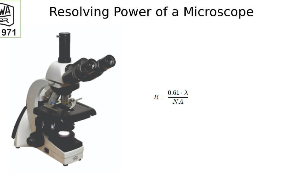
- Lower Resolution: Although PCM can reveal fine cellular structures, it still has limited spatial resolution compared to more advanced techniques like electron microscopy or super-resolution microscopy. For extremely detailed imaging, these higher-resolution techniques may be necessary.
- Optical Setup: Proper alignment of the phase contrast condenser, phase rings, and objective lenses is critical for obtaining high-quality images. Improper setup can lead to distorted or unclear images, which can hinder accurate analysis.
Conclusion: Why PCM Matters
Phase contrast microscopy is a powerful and versatile tool that enables researchers to study living cells, tissues, and microorganisms in their natural, unstained state. Its ability to provide high-contrast, real-time images without damaging the sample makes it indispensable in cell biology, microbiology, developmental biology, and medical diagnostics. While there are some limitations, such as halo artifacts and lower resolution, its benefits far outweigh the drawbacks, making it a key technique in biological research.
As technology continues to evolve, PCM will remain a vital tool for observing the dynamic processes that drive life at the cellular level and KYOWA-GETNER microscopes can be your partners in research. Whether you’re studying cell function, disease progression, or microbial behavior, phase contrast microscopy is an essential technique that can provide valuable insights into the world of living organisms.


